
Preventing Contamination in Tissue Culture Facilities
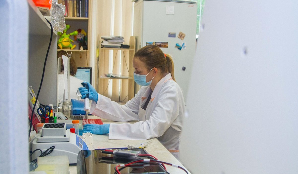
Contamination rarely arrives without warning
Anyone who has watched a flask of carefully nurtured cells turn cloudy overnight knows the particular sinking feeling that follows. Bacterial contamination announces itself with turbidity and a sharp drop in pH; fungal contamination sends fine filaments drifting through the medium; mycoplasma settles in silently and distorts results for months before anyone notices. The good news is that almost every contamination event in a tissue culture facility traces back to a small number of avoidable practices. Getting those right is far easier than trying to rescue an experiment that has already been compromised.
What follows is a practical look at the habits, routines and monitoring that keep cultures clean, whether you run a small academic lab, a biotech R&D suite or a clinical diagnostics service handling primary specimens.
Cleanroom behaviour: the human factor
People are the single largest source of contamination. Every operator sheds particles, and a single careless movement can carry airborne organisms straight into an open flask. Aseptic technique is not a formality; it is the work.
- Dress properly before you enter. Clean lab coat, gloves, tied-back hair, no dangling jewellery, no nail polish or false nails, and sleeves that do not trail across the work surface.
- Work in the right zone. In a Class II cabinet, keep materials at least 10 cm inside the front edge and never let anything pass between the HEPA filter and your open vessel. Nothing should sit downstream of a culture.
- Move slowly and talk sparingly. Rapid arm movements disrupt the laminar curtain, and speaking or coughing near an open plate sends a plume of organisms exactly where you do not want them.
- Clean in the right direction. Wipe surfaces with 70% isopropyl alcohol or ethanol using unidirectional strokes, working from the cleanest area outwards. Allow the alcohol to evaporate fully; it is the wet contact time and the subsequent evaporation that matter, not a quick swipe.
- Use the cabinet correctly. UV lamps are for idle periods only, never while you are working, and glass or plastic in the airflow path should be kept to a minimum.
Cabinet airflow should be verified and serviced on a defined schedule, with records kept alongside your other equipment logs.
Incubator hygiene: the quiet culprit
Incubators are warm, humid and full of nutrients, which makes them excellent culture vessels in their own right. Water trays are the usual offender, particularly in humidified CO2 incubators.
- Use sterile distilled water in humidifying trays, and change it weekly rather than topping it up. Topping up concentrates contaminants.
- Clean on a rota. Empty, wash and disinfect each incubator on a documented cycle, typically monthly for busy facilities, and always after any contamination event.
- Do not overcrowd. Airflow and temperature stability suffer when shelves are packed, and plates near the door experience more fluctuation.
- Monitor independently. Use a separate thermometer and CO2 logger so you can detect a drift before it becomes visible in your cultures.
- Seal long-term cultures. Loosely wrapped plates or sealed flasks reduce the chance of airborne ingress during extended experiments.
Antibiotics: a crutch, not a cure
Penicillin-streptomycin, gentamicin and amphotericin B all have their place, but that place is narrower than many protocols suggest. Antibiotics suppress rather than eliminate contaminants, and low-level bacterial or mycoplasma presence can persist indefinitely beneath the surface, altering cell metabolism, growth rate and gene expression. That is a serious problem when your downstream work is clinical or regulatory.
Reserve antibiotics for short-term protection after primary isolation or during a genuinely necessary transport step, and withdraw them as soon as the culture is established. Critically, routine antibiotic use masks poor technique and breeds resistance. A culture that only survives because of antimicrobials is not a clean culture; it is a suppressed one. Mycoplasma, in particular, is unaffected by most standard antibiotic cocktails, so its absence must be confirmed by testing, not assumed.
Routine monitoring that actually catches problems
Good monitoring is unglamorous, frequent and recorded. It works because it detects problems at the point where they are still cheap to fix.
- Inspect every flask daily. Look for unexpected turbidity, a yellow shift in phenol red, unusual debris or a change in growth pattern. Train everyone to do this, not just senior staff.
- Run negative controls. Incubate an unseeded flask of your complete medium alongside experiments to confirm sterility of media, serum and supplements.
- Use settle plates and surface swabs periodically around the cabinet, incubators and bench areas to build a picture of your facility's baseline.
- Test for mycoplasma on a schedule. PCR-based methods or DNA staining should be part of routine quality control, with results logged and retained.
- Authenticate your cell lines. STR profiling catches cross-contamination between lines, which is often mistaken for a contamination problem when it is actually a labelling or handling error.
When something does go wrong
If contamination is confirmed, act decisively. Quarantine the affected vessel, autoclave and discard it, and disinfect the incubator and cabinet before returning to work. Resist the temptation to salvage a precious flask with a heavy antibiotic dose; in almost every case, restarting from a validated frozen stock is faster, cleaner and more scientifically defensible. Investigate the cause calmly and without blame, review your logs, and adjust the practice that allowed it through. A facility that treats contamination as a systems problem rather than a personal failing is a facility that stays clean.


Aseptic technique protects samples and staff from contamination. Learn hand hygiene, sterile tools and laminar flow cabinet routines.
Biomarker tests help clinicians detect disease and monitor treatment. This article explains common assays, sample handling and result interpretation.
CRISPR gene editing offers powerful tools for research. Discover experimental design, delivery methods and ethical approval steps in UK institutions.
Effective inventory systems reduce waste and keep experiments running. Learn labelling, stock rotation and supplier negotiation strategies for busy labs.